A liver cell can keep working for years, but many of its proteins and organelles cannot. Enzymes become damaged, mitochondria lose efficiency, and bits of membrane reach the end of their useful lives. The cell cannot simply let this material accumulate. Much of it eventually reaches a lysosome, an acidic compartment where large biological molecules are taken apart and their building blocks made available again. That process is more selective and organized than the familiar description of lysosomes as cellular rubbish bins suggests.
What is inside a lysosome?
A lysosome is enclosed by a membrane that separates dozens of digestive enzymes from the cytoplasm. Its enzymes include proteases, which cut proteins into smaller pieces, lipases that act on lipids, and nucleases that break down nucleic acids. Each enzyme acts on particular chemical bonds. Together they can disassemble a remarkably diverse mixture of biological material, but no single enzyme does every job. In a microscope, lysosomes are not all perfect spheres of identical size. Their appearance depends on what they contain and on where they are in the traffic of material moving through the cell.
The lysosomal interior is acidic, typically near pH 5 rather than the approximately neutral conditions of the cytosol. A membrane protein called a V-type ATPase uses energy to move protons inward. This acidity helps many lysosomal enzymes work efficiently. It also creates a degree of separation between ordinary cytoplasmic chemistry and digestion. The lysosomal membrane is not an indestructible wall, and damage to it can have serious consequences; nevertheless, compartmentalization makes it possible to dismantle old components without dismantling the whole cell.
How unwanted material gets there
There is no single entrance into the lysosomal system. Material taken up from outside a cell commonly travels through endosomes, compartments that sort incoming cargo. Some receptors are returned to the cell surface for another round of use, while other cargo continues through the endosomal pathway toward degradation. Immune cells can engulf particles or microbes into larger vesicles, which subsequently interact with lysosomes. Inside the cell, obsolete components arrive by several forms of autophagy. These routes converge on digestion, but they start with different decisions about what is worth retaining.
Consider a receptor that has brought cholesterol-containing particles into a cell. A membrane-bound sorting system can return the receptor for further use while directing the particle’s contents toward processing. Recycling the receptor matters: manufacturing a new protein every time would be wasteful. This is one reason membrane traffic and lysosomal function are tightly linked. The destination of a molecule depends on sorting signals, the machinery that recognizes them, and the needs of the cell at that moment.
Three routes into the lysosomal system:
- Endocytosis: extracellular molecules enter through the plasma membrane and are sorted in endosomes.
- Phagocytosis: specialized cells engulf large particles or microbes for processing.
- Autophagy: cellular material, including damaged organelles, is packaged for delivery from within.
Autophagy: packaging a part for dismantling
During macroautophagy, a membrane grows around a portion of cytoplasm or an organelle, creating a double-membrane structure called an autophagosome. Its contents are not digested immediately. The autophagosome must first mature and fuse with a lysosomal compartment, allowing enzymes to reach the enclosed material. The membrane of the new compartment is remodeled, and the resulting breakdown products can be transported into the cytosol. Autophagy therefore involves recognition, packaging, delivery, fusion, digestion, and recovery—not a single switch that turns recycling on.
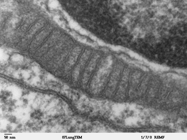
Some forms are relatively broad, capturing a portion of cytoplasm during periods of nutrient stress. Others are selective. Mitophagy, for example, directs particular mitochondria toward removal. Selective pathways depend on molecular tags and receptors that connect the cargo to the machinery forming the autophagosome. The distinction matters because an energetic cell may need to remove a poorly functioning mitochondrion without sacrificing its entire mitochondrial population. Whether a mitochondrion is removed also depends on its physical condition and on wider cellular signaling networks.
What happens to the recovered materials?
Digestion is not the end of the story. Proteins can yield amino acids; nucleic acids yield smaller components; and complex carbohydrates and lipids are reduced to usable molecules. Transporters in the lysosomal membrane move many of these products out for reuse or further metabolism. This recovery becomes particularly important when nutrients are scarce, although lysosomal turnover continues in well-fed cells too. Autophagy is not merely an emergency response. It contributes to housekeeping during ordinary growth, development, and long-lived tissue maintenance.
The lysosome also participates in signaling. Its surface hosts proteins involved in detecting nutrient availability, including components of the mTORC1 signaling network. When circumstances change, this system helps coordinate growth with the availability of resources. That arrangement is logical: a compartment handling recycled nutrients is well placed to participate in decisions about whether the cell should build more material or conserve what it has. It would be inaccurate, however, to describe the lysosome as the cell’s sole metabolic controller. The signal is integrated with information from elsewhere.
When recycling fails
A defect in a single lysosomal enzyme can have consequences far beyond one chemical reaction. In some lysosomal storage disorders, a particular substance cannot be degraded efficiently and accumulates within cells. Different organs may be affected because tissues make and handle different amounts of that substance. Disorders involving trafficking proteins, membrane integrity, or autophagy machinery can also disrupt recycling, even when the individual digestive enzymes are normal. These conditions demonstrate how much of cell maintenance depends on correct delivery as well as chemical breakdown.
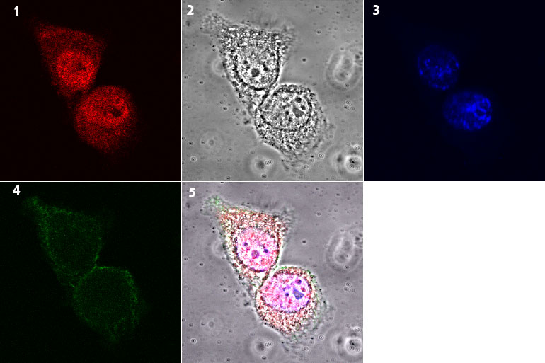
Researchers assess lysosomal function in several ways. Fluorescent probes can report acidity, microscopy can follow particular cargo, and experiments can monitor the movement of proteins that change location during autophagy. One frequently used marker, LC3, associates with autophagosomal membranes, but counting LC3-positive spots alone does not establish how much material is being degraded. A buildup can mean more autophagosomes are being made or that fewer are being cleared. Scientists therefore measure autophagic flux—movement through the entire pathway—rather than treating a static image as proof of activity.
Recycling is not the same as cell death
Autophagy is sometimes described as a form of cellular self-destruction, but ordinary autophagy primarily supports maintenance and survival. A cell can selectively digest a damaged organelle while preserving the rest of its structure and continuing to function. Programmed cell death, including apoptosis, is a different coordinated process in which the entire cell is dismantled and removed. The two systems can influence one another under stress, but seeing increased autophagy-related markers is not by itself proof that a cell is dying.
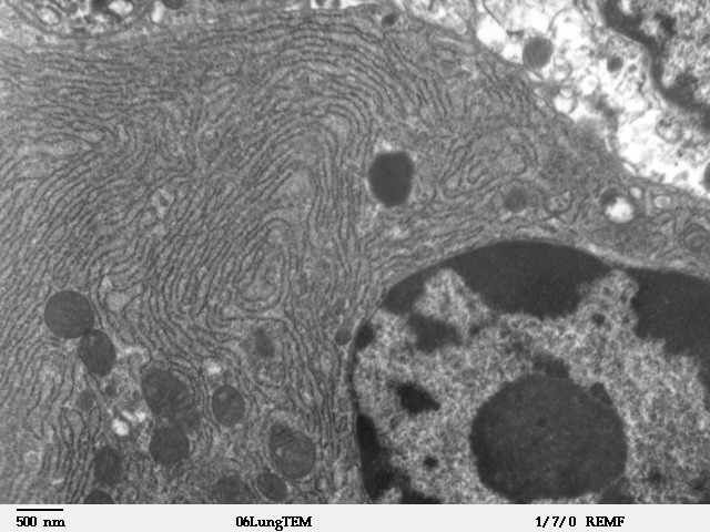
This distinction matters when reading disease research. A tissue with many autophagosomes might be mounting a protective response, experiencing unusually high demand for recycling, or struggling to complete digestion. Researchers need time-resolved evidence of cargo clearance and cell health to distinguish these explanations. A microscope picture of dark intracellular vesicles, however striking, cannot answer all those questions. Mechanism requires measurements of the process, not only images of its structures.
Recycling is part of staying alive
Lysosomes make survival possible through controlled destruction. Cells continuously manufacture, transport, assemble, and dismantle molecules. Without removal and recovery, even carefully built structures would become an accumulating liability. The useful question is not whether a lysosome destroys things, but how a cell decides what to keep, what to repair, and what to break apart. That connects lysosomal biology to the endoplasmic reticulum, protein quality control, and the wider economics of cellular energy discussed in Why Cells Need ATP.