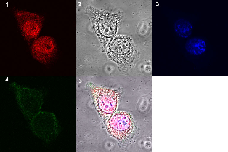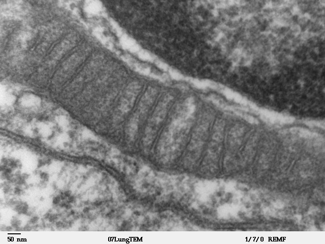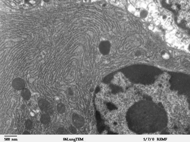
The endoplasmic reticulum, or ER, is often shown as a folded sheet beside the nucleus. It is an easy picture to remember and an incomplete one. In a living cell, the ER is a connected system of sheets and narrow tubules that extends through much of the cytoplasm. Its membranes change shape, meet other organelles and help the cell manage everything from secreted proteins to calcium signals.
Two regions are usually described: rough ER, whose cytosolic surface bears many ribosomes, and smooth ER, which lacks that dense ribosome covering. They are not two unrelated organs. The same continuous system performs different kinds of work in different locations, and its appearance varies enormously from one cell type to another.
A route for proteins leaving the cell
Imagine a pancreatic cell making a protein hormone for secretion. Translation starts on a ribosome in the cytosol. As a signal sequence emerges from the growing polypeptide, a signal recognition particle can recognize it and direct the ribosome–nascent-chain complex toward the ER membrane. At the ER, the ribosome associates with machinery that allows the new polypeptide to enter the ER lumen or become inserted into the membrane while synthesis continues.
This targeting system is selective. Proteins destined to remain in the cytosol generally do not enter the ER, while many secreted proteins, membrane proteins and proteins meant for compartments of the secretory pathway begin processing there. The underlying experiments helped establish the idea that proteins carry sorting information in their sequences. The endoplasmic reticulum chapter of Molecular Biology of the Cell describes the classic microsome experiments that supported the signal hypothesis.
The rough appearance of some ER regions reflects ribosomes temporarily working on ER-targeted proteins. Those ribosomes are not permanently assigned to a separate class. A ribosome that has completed translation can return to the cytosolic pool and translate a different message later. That mobility helps explain why ‘rough’ and ‘smooth’ are descriptions of regions rather than fixed identities.
Folding is part of production
A newly synthesized chain is not automatically a useful protein. It may need to fold into a particular three-dimensional shape, form disulfide bonds, acquire sugar groups or assemble with partner chains. The ER lumen contains enzymes and chaperones that support these processes. Some proteins are retained until they reach an acceptable state, while others are moved onward through the secretory pathway.
Many secreted proteins acquire N-linked carbohydrate chains in the ER. Those sugars are not merely decorations; they can participate in folding surveillance and later biological recognition. A protein that repeatedly fails quality-control checks may be directed toward ER-associated degradation, often abbreviated ERAD. In this route, unsuitable proteins are moved toward the cytosol and targeted for breakdown, commonly through the proteasome system.
Quality control protects the cell against allowing too many faulty proteins to proceed. It also makes protein secretion resource-intensive. A cell producing large amounts of antibody or digestive enzyme has a different ER workload from a cell making only modest quantities of secreted protein. The size and organization of the rough ER often reflect those demands.
The unfolded protein response is not simply a failure alarm
When the production and folding of proteins become badly mismatched, unfolded or misfolded proteins may accumulate in the ER. Cells respond through signaling pathways collectively called the unfolded protein response, or UPR. These pathways can temporarily reduce the influx of newly made proteins, increase the capacity for folding and degradation, and change gene expression.
A short-lived response can help restore balance; it is not automatically evidence that the cell is about to die. A prolonged, severe disruption can have harmful consequences, but the outcome depends on the cause, tissue and duration of the stress. It is important not to collapse every observation of ‘ER stress’ into a single diagnosis or mechanism.
Research papers sometimes track proteins associated with UPR pathways as indicators. Those indicators need interpretation alongside experimental conditions, because they can change for more than one reason. The pathway under study and the measurement chosen should be made explicit before drawing conclusions about cell health.
Why smooth ER matters
Smooth ER regions support the synthesis and processing of lipids that become components of cellular membranes. Membrane expansion is not a trivial housekeeping task: every cell division and much of organelle maintenance require appropriate mixtures of phospholipids and other lipid molecules. The ER is a central location for producing many of these building blocks and distributing them to other membranes.
In some tissues, smooth ER is especially developed because the cell performs specialized chemistry. Cells involved in steroid hormone synthesis, for instance, rely on several membrane-associated enzymatic steps that involve the ER as well as mitochondria. Liver cells carry out metabolic transformations involving ER-associated enzymes, although the familiar phrase ‘the ER detoxifies chemicals’ is much too broad to describe a single universal pathway.
Different smooth ER structures have different jobs. The distinction is clearest in muscle, where a specialized ER network called the sarcoplasmic reticulum helps store and release calcium. Calcium movement in this system regulates muscle contraction and relaxation. That is a functional specialization, not evidence that every smooth-ER tubule operates like a muscle calcium store.
The calcium compartment
Calcium ions act as signals inside many cell types. Their concentration in the cytosol is often kept relatively low compared with certain intracellular stores, enabling small releases to produce meaningful changes in signaling. ATP-dependent pumps move calcium into the ER, while channels can release it in response to appropriate cues.
In muscle, tightly coordinated calcium release allows contractile proteins to interact, and reuptake allows the fiber to relax. In other cell types, calcium signals contribute to secretion, metabolism and gene regulation. Because the timing and location of a calcium signal matter, merely measuring total calcium in a cell may obscure the event a researcher wants to study.
This is one reason cellular compartments are indispensable. The ER does not need to act as a sealed box for every molecule, but its membrane creates distinct chemical spaces that can be regulated by specialized proteins.
How the ER passes material to the Golgi
Proteins that pass ER quality control may leave at specialized exit sites, commonly in transport carriers associated with COPII coat proteins. These carriers participate in traffic toward the Golgi apparatus, where proteins can undergo further modification and sorting. Retrieval systems also return proteins and trafficking machinery that belong in the ER.
It is tempting to describe the process as a straight conveyor belt: ER, Golgi, cell surface. Actual transport is more regulated. Some proteins are retained, others are retrieved, and membrane traffic must remain balanced as materials enter and leave each compartment. Studying this process means following cargo movement, not just inspecting static pictures of organelles.
The ribosome’s role at the start of this pathway is covered separately in Inside the Cell’s Protein Factories. Protein synthesis and protein delivery are connected, but they are distinct problems for the cell to solve.
What microsomes taught biologists
A useful example comes from a laboratory preparation rather than a living-cell photograph. When cells are disrupted, portions of the ER membrane can reseal into small vesicles called microsomes. Some retain ribosomes and can reproduce parts of ER protein translocation in an experimental system. By comparing translation reactions with and without microsomes, researchers learned how signal sequences can direct a newly synthesized protein into a membrane-bound compartment.
The experiment helped distinguish two models. In one, proteins would be made fully in the cytosol and somehow pushed into an organelle afterward. In another, certain polypeptides would be directed toward the ER while translation was still underway. The presence of an ER-derived membrane preparation altered the form and processing of selected proteins, supporting the latter mechanism for many secreted proteins.
This does not mean every protein enters organelles the same way. Mitochondria, nuclei and peroxisomes use different combinations of targeting sequences and transport machinery. The importance of the ER story is that the cell can read destination information from a protein’s sequence and act on it during synthesis.
A dynamic network rather than a background organelle
Modern cell imaging shows that ER membranes make extensive contacts with mitochondria, endosomes and the plasma membrane. Such contact sites can support lipid exchange and signaling without merging the two organelles into one compartment. Meanwhile, the balance between sheets and tubules changes with protein composition and cellular activity.
It is useful to connect this to Why Cells Need ATP: folding reactions, membrane transport and calcium pumping all draw on organized cellular energy systems. The ER’s ability to serve different roles depends on the right proteins being available in the right locations, with adequate energy and precise regulation.
The question to ask of any ER diagram is therefore not only ‘What does this organelle look like?’ but ‘Which activity is being shown?’ A smooth tubule, a ribosome-covered sheet and an ER contact site may belong to the same network while revealing very different aspects of its work.
References
- Alberts B et al. The Endoplasmic Reticulum. Molecular Biology of the Cell. Protein targeting, rough and smooth ER, microsomes and secretory processing.
- Alberts B et al. From RNA to Protein. Molecular Biology of the Cell. Ribosome behavior and translation.
- Alberts B et al. The Mitochondrion. Molecular Biology of the Cell. Intracellular membranes and energy-linked transport context.
Featured image: Louisa Howard, Public domain. Actual cell organelles; original 640×480