Many reactions that support life are energetically possible but painfully slow without a catalyst. Enzymes help those reactions proceed fast enough to matter. They can bind substrates, orient reacting groups and stabilize transition states, but they do not overturn the laws of thermodynamics. Measuring their effects also takes care: rates depend on concentrations, temperature, pH and the assay used.
A chemical obstacle
Many reactions that are thermodynamically possible happen too slowly to support a living cell. An enzyme speeds a reaction by providing a pathway with a lower activation free energy. It is not consumed in the net reaction and does not make an unfavorable equilibrium favorable by itself.
How the active site works
An enzyme usually has a region called the active site that binds substrates and helps the chemical reaction occur. The active site is not always a rigid lock. Binding can shift the shapes of both enzyme and substrate, and several amino-acid side chains may participate in catalysis. Some enzymes position reacting groups precisely, stabilize charge developing in a transition state or briefly form covalent intermediates. Metal ions and non-protein cofactors are also essential for many enzyme reactions. The mechanism can differ dramatically even between enzymes that transform similar molecules. Describing every enzyme as “grabbing a substrate and snapping it apart” is therefore a poor general explanation of enzyme chemistry.
Substrate concentration and saturation
At low substrate concentration, adding more substrate often increases the rate because active sites are less frequently occupied. For many simple enzyme systems, rate rises toward a maximum as the available enzyme approaches saturation. The familiar Michaelis–Menten equation describes a useful model with specific assumptions, not a universal law for every enzyme or experimental setting. The parameter Km has a precise mathematical definition within that model and should not automatically be called binding affinity. Real enzymes can have several substrates, multiple regulatory sites or complicated reaction sequences. Researchers fit kinetic models to measurements and check whether the assumptions are reasonable before interpreting reported constants.
Temperature and pH shape activity
Increasing temperature can speed molecular movement and initially increase some reaction rates, but excessive heat may destabilize the enzyme. The resulting activity curve depends on enzyme stability, assay duration and other conditions. pH affects the protonation of chemical groups involved in binding and catalysis, so different enzymes work well in different environments. Digestive enzymes functioning in acidic stomach contents face different conditions from enzymes in the cytosol. These differences reflect molecular adaptation, not an inherent property that one pH is ideal for life as a whole. Experiments reporting an optimum must describe the measurement conditions rather than presenting the number as fixed for all contexts.
Inhibitors and regulation
Molecules can inhibit enzyme activity through several mechanisms. Some compete with a substrate for access to the active site, while others bind at separate sites or react with essential enzyme groups. Reversible and irreversible inhibition should be distinguished, as should the precise kinetic behavior observed under defined conditions. Cells also regulate enzymes by adding phosphate groups, changing protein partners or altering the amount of enzyme present. Feedback inhibition can restrain a pathway when enough of a product accumulates. Many medicines act through enzyme targets, but a simple diagram of an inhibitor blocking an active site rarely captures the complete pharmacology, including distribution, metabolism and effects on other proteins.
Enzymes in industrial and medical work
Enzymes are widely used outside living cells, including in food processing, detergents, biotechnology and analytical tests. Their specificity can enable reactions under milder conditions than some conventional chemical processes, but stability and cost still matter. Laboratories measure enzyme activity to characterize samples, and certain blood tests detect activities associated with tissue injury or organ function. A measured activity is not always the same as the amount of enzyme present, and it cannot by itself determine a diagnosis. In research, scientists investigate enzyme mechanism with structural studies, isotope experiments and kinetic measurements. Together these approaches reveal how a protein guides chemistry, rather than merely showing that a reaction runs faster.
Why a measured reaction rate needs controls
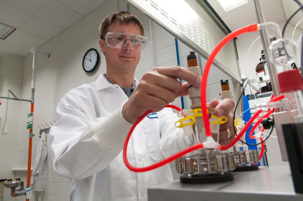
Imagine following catalase activity by collecting oxygen released as hydrogen peroxide is broken down. The volume of gas alone is not an enzyme-rate measurement until the experiment accounts for time, substrate amount, temperature and the possibility of gas escaping from the apparatus. A useful comparison varies one factor while holding others as constant as possible. As hydrogen peroxide concentration rises, the early reaction rate may increase, but the simple proportional relationship need not continue indefinitely because active sites can become occupied and high substrate concentrations can cause other complications. A straight line through every data point should not be expected. The OpenStax discussion of enzyme kinetics and regulation provides a framework for interpreting substrate availability, active-site behavior and inhibition without confusing the amount of enzyme with its catalytic efficiency.
Comparing enzyme activity across samples
When scientists compare enzyme samples, they usually report activity relative to a defined amount of material. A reaction that consumes 10 micromoles of substrate in one minute is not necessarily twice as efficient as one that consumes five: the first tube may contain twice as much enzyme. Specific activity—often expressed as activity per milligram of protein—provides another comparison, although the protein measurement must itself be reliable.
Rates should be measured over an interval when the product signal is approximately linear with time. If most substrate has already been used or product inhibits the enzyme, a late measurement may underestimate initial activity. A blank without active enzyme can reveal background changes in absorbance or fluorescence. Stating assay temperature, pH, substrate amount and normalization method makes reported numbers interpretable rather than merely impressive.
Why a structure alone is not the answer
A detailed enzyme structure can suggest which residues interact with a substrate, but it does not automatically establish a reaction mechanism. The protein moves during operation, water molecules can participate, and the most important transition-state geometry may never be captured directly in a static model. To test a proposed mechanism, scientists may replace one amino acid, examine how reaction rates change, measure isotope effects or compare related substrates. A single altered residue can affect stability as well as catalysis, complicating interpretation. Combining structural and kinetic evidence makes a stronger account than inferring chemistry from an attractive molecular picture.
Use catalase to make the mechanism concrete
Catalase helps decompose hydrogen peroxide into water and oxygen. Hydrogen peroxide is a reactive by-product of some cellular processes, so converting it efficiently can protect cells from damage. The enzyme does not change the chemical identities of the starting and ending substances or shift the reaction’s equilibrium; it offers a faster route by stabilizing intermediate arrangements along the reaction pathway. A demonstration using bubbles from peroxide and tissue can show that catalysis occurs, but bubble volume alone is a poor quantitative measure of enzyme kinetics without controlled concentrations, temperature and geometry. [1]
What an inhibitor changes in a rate curve
Consider an enzyme tested at several substrate concentrations, first on its own and then in the presence of a potential inhibitor. Measuring only one point is rarely enough to identify the mechanism. Under a simple reversible competitive-inhibition model, the apparent substrate concentration needed for a given fractional rate increases, while the limiting rate at sufficiently high substrate can remain similar. Other mechanisms produce different patterns.
Real enzymes do not always obey a simple Michaelis–Menten model: some have multiple interacting subunits, show cooperativity or lose activity during the assay. Researchers compare models with replicate measurements and residuals rather than assigning a mechanism solely by how a plotted line looks. A fitted parameter summarizes the observations under stated conditions; it is not an intrinsic universal constant for every laboratory setup.
References
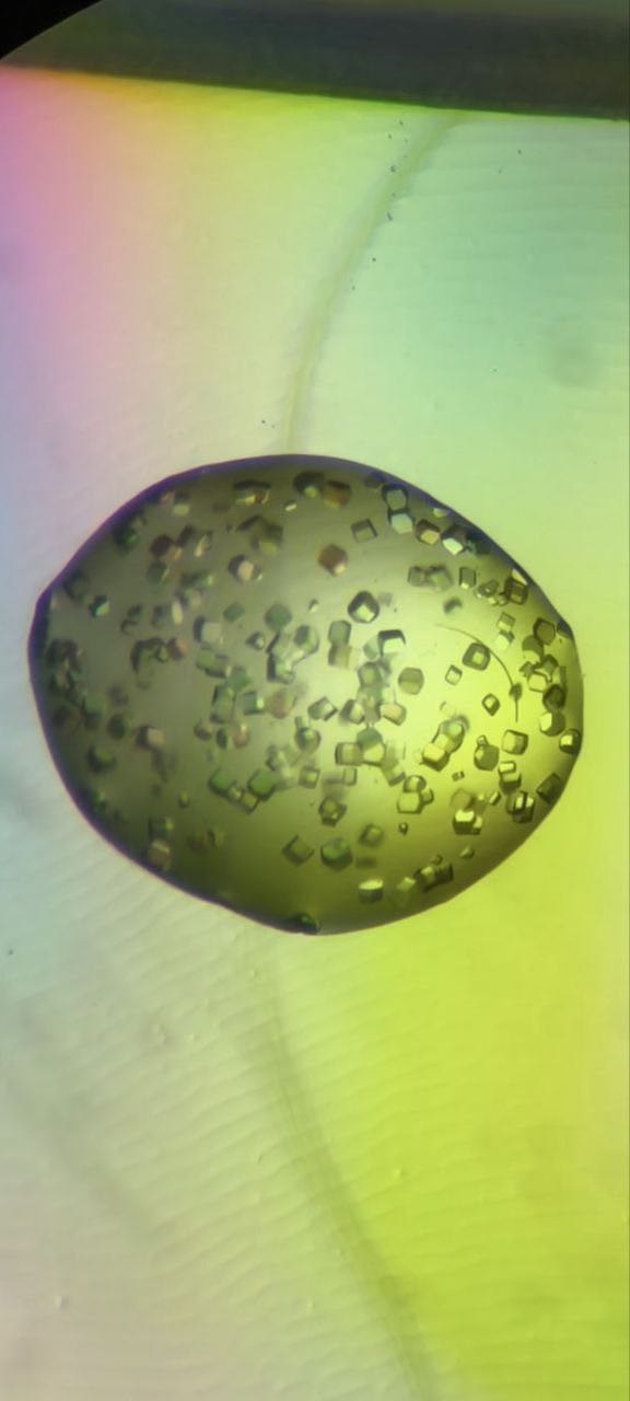
Featured image: Ilyassova Bayan, CC0 1.0. Real lysozyme crystals; portrait source crop may be needed