When you decide to lift a cup, the muscles involved do not receive a single continuous electrical current from the brain. Nerve cells generate brief voltage changes, pass signals across chemical junctions and activate groups of muscle fibers. Each step depends on ions moving through specialized membrane proteins. The sequence is fast, coordinated and vulnerable to different kinds of disruption.
A difference across a membrane
Nerve cells maintain unequal concentrations of ions across their membranes. Ion channels and transporters help establish a resting membrane potential. This voltage is not an electrical current flowing freely through the body; it comes from ion movement across and along living cell membranes.
Timing and coordination
Movement depends on more than one nerve firing. Networks in the brain and spinal cord integrate sensory information and coordinate many muscles. Myelin can speed conduction along certain axons, but the speed and pattern of signaling differ widely among cell types.
How an action potential begins
A sufficiently strong change in membrane voltage can cause voltage-gated sodium channels to open rapidly. Sodium entering the cell shifts the voltage toward positive values, which promotes further channel opening. Shortly afterward, sodium channels inactivate and potassium channels help return the membrane potential toward its resting level. This rapid sequence forms an action potential. Many neurons use action potentials as long-distance signals, although different nerve and muscle cells have distinct channel combinations and electrical behavior. A small stimulus that does not reach threshold may produce a local voltage change without launching a full action potential. The threshold mechanism helps separate graded local inputs from all-or-none spikes that travel along an axon.
Why the signal travels in one direction
When a region of an axon depolarizes, local current influences adjacent membrane, bringing neighboring voltage-gated channels toward activation. The region that just fired is briefly less able to fire again because sodium channels are inactivated. This refractory period contributes to orderly propagation. In myelinated axons, insulating myelin wraps reduce current loss, and action potentials are renewed at gaps called nodes of Ranvier. This organization supports rapid conduction without requiring every segment of membrane to generate a full spike. Loss or damage of myelin can disrupt conduction, but the effects depend on which nerves are affected and the underlying cause. Speed also depends on axon diameter and other cellular properties.
A message crosses the synapse
At many chemical synapses, an arriving action potential opens voltage-gated calcium channels in a nerve terminal. Calcium entry triggers vesicles to release neurotransmitter molecules into the narrow synaptic cleft. Those molecules bind receptors on the next cell and alter its behavior. Some receptor responses make a neuron more likely to fire, while others make it less likely. A single neuron receives many such influences at once and integrates them across time and space. The common classroom picture of one nerve cell directly commanding another overlooks this constant balancing of inputs. Electrical synapses also exist, allowing current to pass through specialized connections, but chemical synapses are central to much nervous-system signaling.
From a motor neuron to muscle
Why nerve conduction and muscle activation are separate steps
An electrical impulse reaching a motor-neuron terminal does not jump directly into a muscle fiber as an uninterrupted current. Arrival at the terminal opens voltage-gated calcium channels; incoming calcium promotes release of acetylcholine. The transmitter crosses a short extracellular gap and activates receptors at the motor end plate. If depolarization reaches threshold, the muscle membrane generates its own electrical response. That response spreads along the membrane and into transverse tubules, coupling electrical activity to calcium release inside the fiber. Calcium then permits actin and myosin to interact. Drugs or disease can interfere with any one of these stages, which is why normal nerve conduction does not by itself guarantee normal force production. Recording muscle activity is informative, but a measured signal is not the same as a complete assessment of strength, coordination or sensation.
At the neuromuscular junction, a motor neuron releases acetylcholine, which activates receptors on a skeletal muscle fiber. The resulting electrical response can trigger a muscle action potential that travels along the membrane and into transverse tubules. This signal activates mechanisms that release calcium from the sarcoplasmic reticulum. Calcium allows contractile proteins to interact, producing force through repeated molecular cycles that consume ATP. Relaxation requires calcium to be removed from the cytosol and for cross-bridge cycling to decline. The full process links an event lasting milliseconds to the coordinated movement of an arm or leg. Voluntary movement also depends on motor circuits, sensory feedback and many muscles working together.
Signals can fail in different ways
Weakness or abnormal sensation can result from problems at different levels: nerve pathways, myelin, synapses, ion channels, muscles or circulation. Similar outward symptoms do not imply identical mechanisms. Researchers and clinicians use electrical recordings and other tests to study where signals are altered. For example, electromyography measures electrical activity associated with muscle activation; it does not directly photograph a nerve impulse. Ion channels are also targets of many medicines and toxins, which is one reason physiology is useful beyond the classroom. A general explanation of action potentials cannot diagnose a personal symptom. The central principle remains that electrical signaling depends on carefully maintained ion gradients and precisely timed changes in membrane permeability.
Follow one command to a muscle
A motor command eventually reaches a motor neuron whose axon conducts an action potential toward a muscle. At the neuromuscular junction, the arriving signal opens voltage-gated calcium channels in the nerve terminal. Calcium entry helps trigger release of acetylcholine, which binds receptors on the muscle membrane. If the resulting depolarization reaches threshold, the muscle fiber produces its own electrical signal and begins the sequence that releases calcium for contraction. The neural impulse does not travel directly into the muscle unchanged: information crosses a chemical synapse and then becomes an electrical response again. [1]
Why a recording is not the whole explanation
An electromyogram records electrical activity associated with muscle activation, but its shape depends on electrode placement, how many motor units contribute and the tissue between electrodes and fibers. It cannot by itself identify a single molecular defect. Similarly, an action-potential diagram is a simplified trace of membrane voltage, not an image of ions moving along a wire. Sodium and potassium conductances change with time and across membrane regions. Measurements of individual channels, nerve conduction and muscle activity address different levels of the same biological pathway.
Restoring ion gradients and cycling muscle proteins require metabolic energy. The molecular source of that work is covered in Why Cells Need ATP.
References
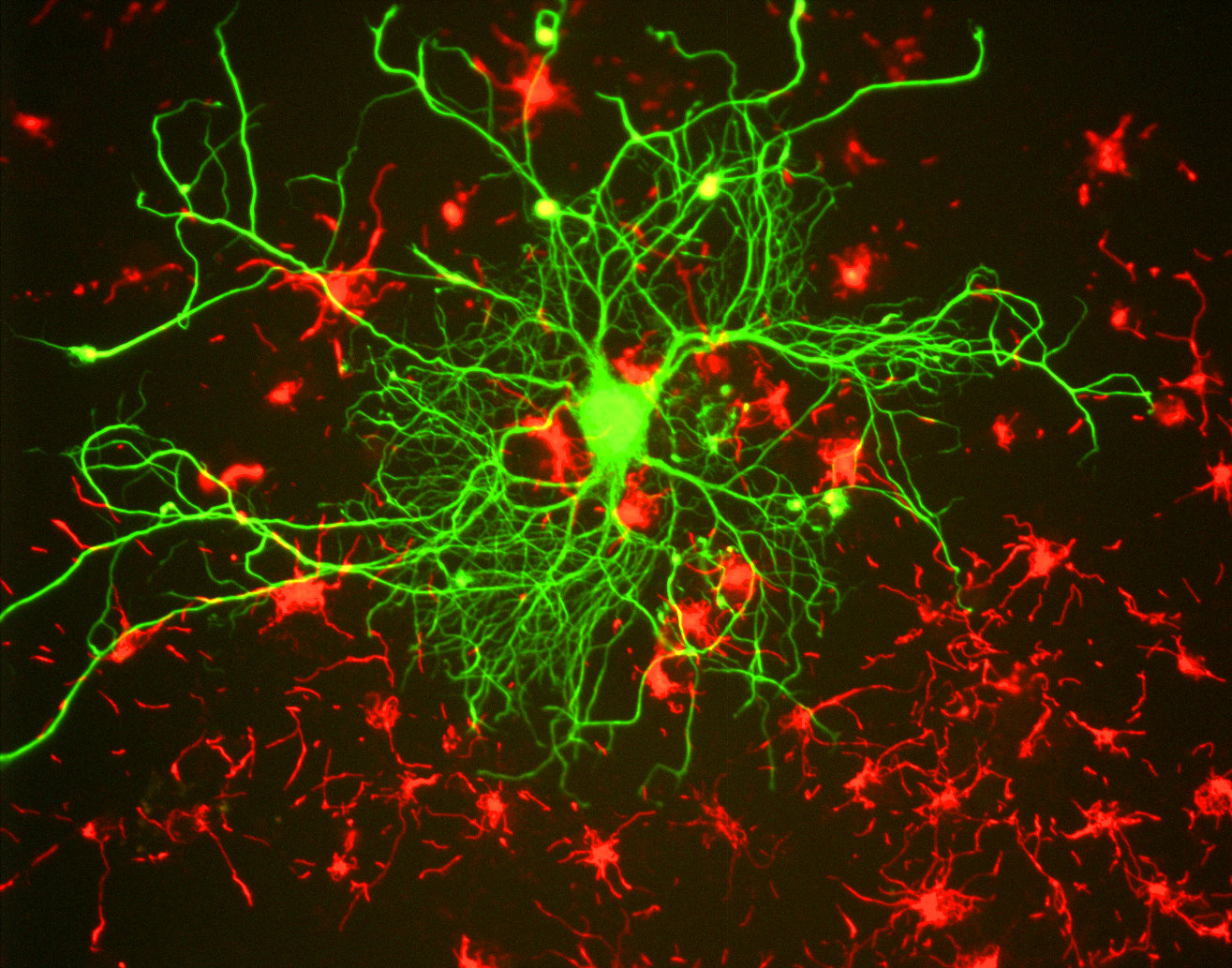
Featured image: GerryShaw, CC BY-SA 3.0. Neuron image, not direct measurement of an electrical impulse