Follow the route of one red blood cell and the heart makes more sense. Blood passes through four chambers and two circulations, with valves responding to pressure differences as the heart fills and contracts. The pulmonary circuit sends blood through the lungs; the systemic circuit carries it to the rest of the body. The routes are connected but not interchangeable.
Two linked circulations
The right side of the heart receives blood returning from the body and pumps it toward the lungs. The left side receives blood from the lungs and sends it into the systemic circulation. These circuits are connected, and in a healthy adult their average flows must match over time.
Valves maintain direction
The tricuspid, pulmonary, mitral and aortic valves respond to pressure differences as the heart contracts and relaxes. They do not actively pull themselves open. Their structure helps prevent significant backflow when the pressure changes.
Electrical signaling in another tissue is examined in The Electrical Signals That Keep Your Body Moving.
Pressure opens and closes valves
Heart valves do not open because a small motor pulls them apart. They respond largely to pressure differences across their leaflets. When pressure in an atrium exceeds pressure in its ventricle, the atrioventricular valve opens and blood can flow forward. As ventricular pressure rises during contraction, that valve closes; once ventricular pressure exceeds arterial pressure, the semilunar valve can open. Valves limit reverse flow and help the chambers pump in an efficient sequence. The sounds heard with a stethoscope are associated primarily with valve closure and related vibrations, not simply with blood splashing through open chambers. Abnormal valve structure or function can change these sounds, but interpreting them requires clinical assessment.
The rhythm is electrical
The sinoatrial node contains specialized cells that normally initiate the electrical rhythm of the heart. Electrical activity spreads through atrial tissue, passes through the atrioventricular conduction system and travels through pathways that coordinate ventricular contraction. The atrioventricular node introduces a delay that helps allow ventricular filling before the major ventricular contraction. Heart muscle cells then translate changes in membrane voltage into calcium-dependent contraction. The timing is essential: if every chamber contracted independently, the paired pumps would be much less effective. An electrocardiogram records summed electrical activity from the body surface; it does not directly measure how much blood the heart pumps with each beat.
Stroke volume and cardiac output
What the two heart sounds actually mark
The first normal heart sound is associated primarily with closure of the atrioventricular valves near the start of ventricular systole. The second is associated with closure of the aortic and pulmonary valves as the ventricles begin to relax. These sounds mark valve events; they are not produced by blood simply passing through an open chamber. Between them the pressure in the ventricles rises enough to send blood into the aorta and pulmonary circulation. The sequence also explains why the ventricles briefly contract with all valves closed before ejection begins. A detailed account of the cardiac cycle connects valve positions, pressure and electrocardiogram timing. For an introductory reader, following the pressure changes is more informative than memorizing four chamber names in isolation.
A heartbeat ejects a certain volume of blood called stroke volume. Cardiac output is the product of stroke volume and heart rate, so changes in either can affect circulation. The amount ejected depends on ventricular filling, resistance to ejection and the muscle’s ability to contract. During exercise, cardiac output ordinarily rises to meet increased demand from tissues. Blood pressure and blood flow are related but not interchangeable measurements. The cardiovascular system regulates vessel diameter, circulating volume and heart activity to maintain delivery of oxygen and nutrients. Any single number, including resting pulse rate, gives only a partial picture of cardiovascular function and must be interpreted in context.
The heart feeds itself
Although the heart contains blood in its chambers, the muscle of the heart receives most of its oxygen through coronary arteries branching from the aorta. The coronary circulation must supply working muscle continuously as demand changes. Restricted supply can impair function, which is one reason coronary circulation matters in cardiovascular disease. Heart valves, chambers and vessels also develop and age in ways that vary among individuals. Anatomical illustrations should show the correct chamber connections and distinguish a schematic from a literal view of the body. A good account of blood flow is ultimately about linked pressures, valves, electrical timing and the demands of tissues, not merely arrows on a diagram.
Why flow cannot be understood from color alone
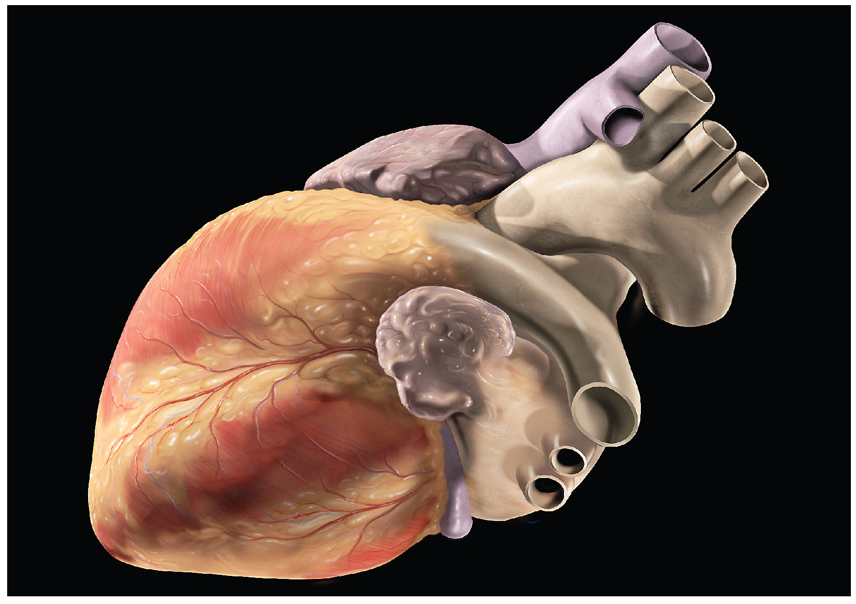
An illustration often colors oxygen-rich blood red and oxygen-poor blood blue. In reality, human blood is red in both states; the shade differs with oxygenation, and blue is a diagram convention rather than a true color of venous blood. Diagrams may also simplify the heart’s three-dimensional orientation and the way major vessels curve. When studying anatomy, it helps to trace the actual route in order instead of relying only on the colors. A credible figure should label its chambers, valves and vessels accurately, and note when anatomy has been simplified for teaching.
Trace one red blood cell through the circuit
Blood returning from body tissues enters the right atrium through the venae cavae, moves through the tricuspid valve into the right ventricle and leaves through the pulmonary valve into the pulmonary arteries. After gas exchange in lung capillaries, blood returns through pulmonary veins to the left atrium, crosses the mitral valve into the left ventricle and travels through the aortic valve into the aorta. This route explains an often-confusing naming rule: an artery carries blood away from the heart, while a vein carries blood toward it, regardless of the blood’s oxygen level. [1]
How cardiac output responds to changing demand
Cardiac output is the product of heart rate and the volume ejected with each beat. This relationship is useful because the circulation can increase delivery without simply making the heart beat as fast as possible. During physical activity, changes in filling, contraction and vascular resistance influence how much blood the ventricles eject. At rest, the amount pumped into the lungs and the amount pumped to the body must remain closely matched over time, even though the two sides work against different pressures. The left ventricle has a thicker muscular wall because systemic circulation normally requires higher pressure than pulmonary circulation. These facts turn the two-circuit diagram into a working mechanical system. A high heart rate alone does not prove that output is adequate: reduced filling time, valve dysfunction or impaired contraction can alter the relationship. Measurements such as stroke volume are interpreted in context rather than used as universal health scores.
References
Featured image: OpenStax Anatomy and Physiology, CC BY 4.0. Specimen illustrates anatomy, not moving blood