With every breath, air reaches millions of tiny spaces called alveoli. Blood passes through fine capillaries alongside them. Oxygen moves from air into blood, while carbon dioxide moves the opposite way. The process seems effortless when the lungs are healthy, but it depends on several conditions lining up at once: air must reach the alveoli, blood must reach the nearby capillaries, and the gases must cross a very thin boundary along the right pressure gradients.
Following a breath to the alveoli
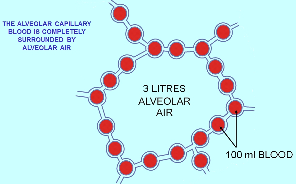
Air enters through the conducting airways and travels down a branching system of bronchi and bronchioles. These passages warm, humidify, and distribute air but are not themselves all sites of substantial gas exchange. The respiratory zone includes the alveoli, whose enormous combined surface area provides room for gas molecules to move between air and blood. At this scale, the lungs resemble a carefully packed interface more than a pair of empty bags. Their architecture increases the contact area while keeping the diffusion distance small.
Alveoli are lined mainly by two classes of epithelial cell. Type I alveolar cells form much of the very thin surface through which gases move. Type II cells produce surfactant, a mixture that reduces surface tension and helps alveoli remain open. An alveolus that collapses or fills with fluid cannot exchange gas as effectively merely because oxygen exists somewhere in the airway. The microscopic structure of the lining, and the condition of the surrounding capillary, are both essential.
What drives oxygen into blood?
Gases move according to differences in their partial pressures. A gas’s partial pressure represents its contribution to the total pressure in a mixture and provides a useful measure of the driving force for diffusion. In a normally functioning lung, the partial pressure of oxygen in alveolar air is higher than in deoxygenated blood arriving through the pulmonary circulation. Oxygen therefore diffuses toward the blood, crosses the respiratory membrane, and enters red blood cells where much of it binds to hemoglobin.
The pressure difference matters more than a vague idea that the blood is ‘hungry’ for oxygen. Hemoglobin binding helps maintain uptake because oxygen entering red cells can be bound, affecting the relationship between dissolved oxygen and total oxygen content. Most oxygen in blood is transported bound to hemoglobin; relatively little is dissolved in plasma. That distinction is why a measurement of oxygen partial pressure is related to, but not identical with, the total amount of oxygen the blood can carry.
Carbon dioxide follows another gradient
Cells generate carbon dioxide during metabolism. Blood returning from tissues transports it to the lungs in several forms: dissolved gas, bicarbonate, and compounds associated with proteins. In the pulmonary capillaries, the relevant reactions shift so that carbon dioxide can move out of the blood into alveolar air. The gradient for carbon dioxide is smaller in pressure terms than for oxygen, but carbon dioxide diffuses effectively through biological membranes under ordinary conditions because of its physical properties.
The bicarbonate system is especially important. In red blood cells, an enzyme called carbonic anhydrase helps interconvert carbon dioxide and water with carbonic acid–related species. Much of the carbon dioxide produced in tissues is thus transported as bicarbonate in plasma. In the lung, the sequence proceeds in the direction that supports carbon dioxide release. Ventilation removes the gas from alveoli, maintaining the gradient for more to diffuse out of the blood.
Ventilation and perfusion must match
Ventilation describes the movement of air into and out of the lungs. Perfusion describes the flow of blood through the pulmonary capillaries. Gas exchange works best when a region receiving air also receives an appropriate blood supply. A poorly ventilated region with continuing blood flow cannot oxygenate that blood adequately. A well-ventilated region with very little blood flow wastes ventilation because there is little blood available for exchange. The relationship is commonly described as ventilation–perfusion matching.
The lungs are not perfectly uniform. Gravity, local vessel behavior, airway dimensions, and other factors create regional differences. Physiological regulation can partially compensate. For example, low alveolar oxygen in a region can cause nearby pulmonary vessels to constrict, redirecting some blood toward better-ventilated areas. This response differs from the more familiar response of many systemic blood vessels to low oxygen. It helps the lungs use their available exchange surface more effectively, although severe or widespread problems cannot be solved by redistribution alone.
| Problem | What is affected? | Why it matters |
|---|---|---|
| Low ventilation relative to blood flow | Fresh gas supply to alveoli | Blood may leave less oxygenated |
| Low perfusion relative to ventilation | Blood available to collect oxygen | Some delivered air contributes less to exchange |
| Thickened exchange barrier | Diffusion distance | Gas transfer can become less efficient |
Why diffusion distance matters
The respiratory membrane consists of closely associated alveolar and capillary structures. Its thinness allows gases to cross rapidly. Fluid accumulating in the alveoli, damage to the capillary barrier, or substantial thickening of the interface can interfere with gas exchange. Loss of effective surface area also matters. A useful conceptual model is Fick’s law: diffusion increases with available surface area and the pressure difference, and decreases as the barrier becomes thicker. The exact physiology is more complex, but these relationships explain many observations.
The law also helps prevent a misconception: faster breathing does not fix every reason oxygen uptake is impaired. If the barrier is damaged, the capillary blood flow is inadequate, or much of the alveolar surface is unavailable, changing ventilation alone may not restore normal exchange. Similarly, a person may be moving air effectively while the blood has too little hemoglobin to transport the usual amount of oxygen to tissues. Air movement, gas diffusion, and oxygen transport are distinct steps.
The role of hemoglobin at tissues
When oxygen-rich blood reaches systemic tissues, the environment differs from that in the lungs. Oxygen is being consumed, local partial pressures are lower, and conditions such as carbon dioxide concentration, pH, and temperature can influence hemoglobin’s affinity for oxygen. Hemoglobin releases oxygen in accordance with these changing conditions. The resulting transfer supports cellular respiration, but it is not equivalent to the cells using oxygen instantly at the point where a red blood cell passes by.
This provides a useful connection to Why Cells Need ATP. Oxygen availability contributes to aerobic metabolism, while ATP-producing pathways themselves operate inside cells. A normal oxygen reading does not reveal every aspect of mitochondrial function. Conversely, cellular energy demand can rise dramatically during exercise, prompting responses in breathing and circulation. The lung, heart, blood, and metabolizing tissue are parts of one coordinated transport system.
How gas exchange is measured
Researchers and clinicians use several measurements to assess different steps. Pulse oximetry estimates the percentage of hemoglobin binding sites occupied by oxygen; it does not directly measure ventilation or total oxygen delivery. Arterial blood-gas analysis can assess oxygen and carbon dioxide partial pressures and acid–base status. Pulmonary function tests characterize airflow and other lung properties, while specialized tests estimate gas-transfer capacity. Each result answers a particular question, and the limitations of the instruments and the patient’s circumstances matter.
The anatomy is also studied with microscopy, imaging, and models that examine airflow and capillary circulation. A single image cannot reveal whether a region has adequate ventilation or perfusion during ordinary breathing. Physiologists therefore combine structural information with measurements taken during activity or controlled changes in conditions. The crucial distinction is between observing the exchange surface and demonstrating that it works as expected.
An exchange that relies on several systems
The alveolus is the final destination of inhaled air, not the final destination of oxygen. Oxygen still has to enter blood, bind hemoglobin, travel through the circulation, leave capillaries, and reach metabolizing cells. Carbon dioxide must make the reverse journey. Lung gas exchange is a vivid example of how a microscopic membrane becomes part of whole-body physiology. For the circulation that carries gases between the lungs and tissues, see How Blood Moves Through the Heart.
References and further reading
Featured image: Cruithne9 · CC BY-SA 4.0.