In the hours before a human cell divides, it faces a copying job with little room for improvisation. The cell must duplicate its chromosomes so that each daughter cell receives a complete genome. The DNA cannot simply be unrolled and photocopied: it is wound around proteins, its two strands point in opposite chemical directions, and the enzymes that make new DNA have strict operating rules. Much of the elegance of replication lies in how the cell works around those restrictions.
DNA copying occurs during the S phase of the cell cycle. The result is normally two double helices, each containing one strand from the original molecule and one newly synthesized strand. This arrangement is called semiconservative replication. It explains both how a sequence can be inherited and why errors during copying can become permanent variants in a cell lineage.
How scientists established the copying principle
The complementary pairing of DNA bases suggested a mechanism long before researchers could watch the machinery work. Adenine normally pairs with thymine, and guanine with cytosine. Separating the two strands would expose a template for constructing the missing partner. But early scientists considered several possibilities: perhaps both original strands remained together, or perhaps old and new DNA became mixed throughout each daughter molecule.
In 1958, Matthew Meselson and Franklin Stahl distinguished these models by growing Escherichia coli in nitrogen containing a heavy isotope, then transferring the cells to medium with a lighter isotope. They separated extracted DNA by density. After one generation, the DNA formed an intermediate-density band; after two, both intermediate and light bands appeared. That pattern was the prediction of semiconservative copying. The experiment did not reveal every enzyme involved, but it provided a physical test of the underlying model rather than relying on the attractive geometry of the double helix alone.
Starting thousands of copies without copying anything twice
A bacterial chromosome often begins replication at one principal origin. Human chromosomes, by contrast, are much larger and generally use many origins. Initiation at multiple sites produces expanding bubbles of copied DNA; two replication forks move outward from each active origin until neighboring regions are completed. Not every potential origin activates in a particular cell cycle, and the timing differs between regions of the genome.
Before S phase, origin-recognition and licensing proteins help load inactive MCM helicase complexes onto DNA. When the cell enters S phase, signaling proteins activate a subset of these prepared origins. The crucial safeguard is that licensing is restricted: an origin that has fired must not begin again before the next cell cycle. Without that control, some chromosome regions would be copied twice while others might be missed. Backup licensed origins can also become useful when nearby forks stall, helping cells complete difficult stretches of DNA.
Starting replication does not mean stripping every chromosome of its packaging. DNA is associated with histone proteins in nucleosomes. The fork must move through that chromatin and restore organized packaging behind it, using a combination of recycled and newly supplied histones. In this sense, replication copies not just a chemical sequence but a chromosome that must remain usable afterward.
The moving replication fork
At the fork, a helicase separates the parental DNA strands. Proteins coat the exposed single strands to keep them available and limit unwanted secondary structures. Ahead of the fork, topoisomerases relieve the twisting stress created as the helix opens. These jobs must happen together: if opening continues without controlling the strain, the DNA becomes difficult to unwind and is more likely to sustain damage.
DNA polymerase reads an existing template and adds the matching nucleotide to a growing strand. Its chemical limitation is important: it can extend DNA only at a free 3′ end, so the new strand grows in the 5′-to-3′ direction. It cannot initiate a completely new strand from nothing. A primase-containing complex must first supply a short starter, or primer, that polymerase can extend.
Each incorporated nucleotide arrives as a deoxyribonucleoside triphosphate. Its chemistry drives formation of the next phosphodiester bond in the sugar-phosphate backbone. The template selects the order of bases, while the polymerase and its partner proteins carry out the reaction. The energy and molecular organization that support the replication machinery connect this process to broader cellular metabolism, discussed in Why Cells Need ATP.
Why one strand is continuous and the other is not
The two template strands are antiparallel. A polymerase moving with the opening fork can extend one daughter strand continuously toward the fork. This is the leading strand. The other daughter strand must still be made 5′ to 3′, so it is assembled in short segments pointing away from the moving fork. This is the lagging strand, and its segments are called Okazaki fragments.
Imagine opening a zipper while two builders work on its exposed halves. One can build steadily in the direction of opening. The other repeatedly starts a short piece farther along and builds backward toward the previous piece. That analogy conveys the geometry, though the real proteins operate in a coordinated molecular complex. On the lagging strand, primase supplies repeated primers; polymerase extends them; enzymes remove the primer material; and DNA ligase seals the remaining breaks in the backbone.
In human cells, polymerase epsilon is strongly associated with leading-strand synthesis and polymerase delta with much of lagging-strand synthesis, while the polymerase-alpha–primase complex helps start new stretches. These assignments describe the main division of labor, not a claim that each enzyme is incapable of working elsewhere. Sliding clamps and clamp-loading factors help keep polymerases engaged with DNA during sustained copying.
What keeps the copy accurate?
Base pairing provides a first level of selectivity, but complementary letters alone do not eliminate mistakes. Many replicative DNA polymerases can proofread: when an incorrect nucleotide is incorporated, a 3′-to-5′ exonuclease activity can remove it before synthesis continues. Additional mismatch-repair pathways inspect newly copied DNA and replace certain mismatched bases that escaped the polymerase.
Accuracy matters because each round of copying creates an opportunity for a sequence change. A persistent mistake can become a mutation after subsequent replication. Yet a cell also experiences DNA damage from oxidation, radiation, or chemical exposure independently of normal replication. Repair systems respond to several kinds of damage; not all changes in DNA originate as polymerase errors, and not every DNA change has a measurable effect on health or function.
A practical distinction is between a copying error, which may alter a base sequence, and a chromosome-level mistake, which may alter the amount or arrangement of whole stretches of DNA. The latter is examined in When Chromosomes Change. Sequence variation and chromosome structure are connected, but scientists use different tests to investigate them.
What happens when a fork stalls?
Replication forks sometimes meet DNA lesions, tightly bound proteins, unusual DNA structures, or shortages of materials needed for synthesis. Rather than simply forcing their way through, cells activate checkpoint responses that can slow the cell cycle and stabilize the fork. Specialized enzymes may help restart synthesis or repair the underlying problem. If damage is severe, a cell may stop dividing or enter a controlled death pathway.
These responses are particularly important in actively dividing tissues. Some anticancer treatments exploit the difficulty rapidly dividing cells face when DNA synthesis or repair is disrupted. But the sensitivity of different cell types and cancers varies; an explanation of replication is not itself a guide to treatment.
Finishing chromosome ends
Human chromosomes are linear, which creates a problem at their ends. Conventional polymerases need primers, and removing the last primer can leave an end that cannot be filled by ordinary synthesis alone. Telomeres are repetitive DNA-protein structures that help protect chromosome ends, while telomerase can extend telomeric repeats in cells where it is active. Telomerase is not equally active in all tissues, and telomere length by itself is not a simple clock that predicts an individual’s age or health.
Replication ends successfully when the cell has duplicated its genome, addressed unresolved copying intermediates, and prepared the sister chromatids for eventual separation. The familiar phrase “DNA makes a copy of itself” hides a coordinated system of initiation, chemistry, error detection, chromatin management, and cell-cycle control. That coordination is what makes the process reliable enough to support the enormous number of cell divisions involved in a human lifetime.
References and further reading
- Molecular Biology of the Cell — DNA Replication Mechanisms, NCBI Bookshelf.
- Molecular Biology of the Cell — Initiation and Completion of DNA Replication, NCBI Bookshelf.
- The Cell — Heredity, Genes, and DNA, including the Meselson–Stahl evidence.
- Origins of DNA replication, research review in Cold Spring Harbor Perspectives in Biology.
- Genomes — Genome Replication, NCBI Bookshelf.
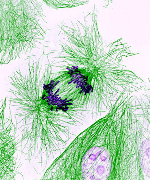
Featured image: National Institutes of Health, Public domain. Shows division, not direct visualization of DNA replication