A chromosome is not a static string of genes. It is a long DNA molecule packaged with proteins, copied before cell division and physically moved into daughter cells. Changes can occur in its number, its internal arrangement, or the amount of DNA it carries. Some have little detectable effect; others disrupt development or contribute to disease. The consequences depend on what changed, where it happened, and which cells carry the change.
It helps to separate two questions from the beginning. Has a cell gained or lost an entire chromosome? Or has a segment within or between chromosomes been rearranged? These are different mechanisms with different laboratory signatures. Neither should be confused automatically with a change in a single DNA letter, which is normally assessed with sequence-based methods.
Why chromosome number matters
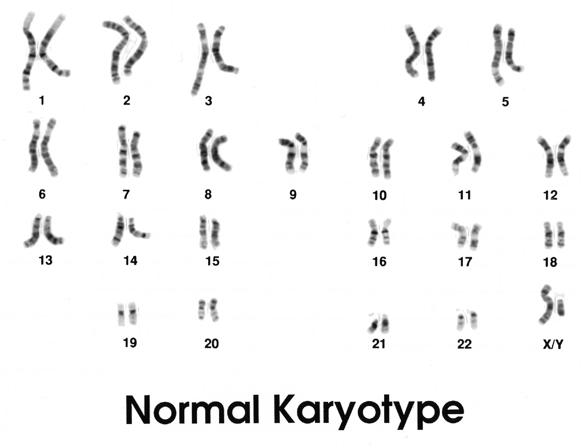
A typical human somatic cell contains 46 chromosomes organized into 23 pairs. Most egg and sperm cells contain 23 individual chromosomes. Fertilization ordinarily restores a pairwise set. A cell with one chromosome too many or too few has a form of aneuploidy, rather than a small change in one gene.
The difference can affect many genes at once. Cells regulate the production of proteins and RNA across entire biochemical networks. Adding an extra chromosome can change the dosage of hundreds of genes, upsetting processes that depend on balanced expression. However, no single chromosome-count rule predicts every effect: the chromosome involved, the developmental stage, and the distribution of affected cells all matter.
Trisomy 21 illustrates the principle. Most people with Down syndrome have three copies of chromosome 21 in their cells. Researchers have linked that extra genetic material to changes in development and an increased likelihood of certain health conditions, but the pattern and severity of features vary among individuals. The chromosome difference does not capture the full person or determine every aspect of their abilities or health.
How an extra chromosome can arise
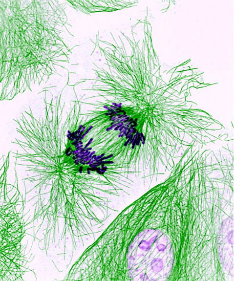
During meiosis, chromosome pairs and then sister chromatids are supposed to separate into reproductive cells. Nondisjunction is a failure of separation that can leave a reproductive cell with an extra or missing chromosome. If such a cell participates in fertilization, the resulting embryo may begin with an atypical chromosome count.
Many numerical chromosome changes arise spontaneously rather than being inherited from a parent who has the same condition. Some chromosome combinations are incompatible with continued development, while others are associated with recognizable conditions. This is one reason that the simple phrase “a mutation passed down through the family” does not fit every chromosome condition.
Errors can also occur after fertilization, during early mitotic divisions. If a change happens after some cells have already formed, the body may contain two or more cellular populations with different chromosome complements. This is called mosaicism. The fraction of affected cells may differ among tissues, which complicates both effects and detection. A blood sample does not necessarily represent every tissue in a mosaic individual.
Deletions and duplications: when the amount of DNA changes
Not all chromosome abnormalities involve whole chromosomes. A deletion removes a segment, while a duplication adds an extra copy of a segment. These are kinds of copy-number change. Their impact depends on their size and on whether the affected stretch contains dosage-sensitive genes or regulatory sequences. A small change can be important if it disrupts a crucial region; a larger change is not automatically more severe in every case.
One documented example is 22q11.2 deletion syndrome. Many affected individuals are missing roughly three million DNA base pairs from one copy of chromosome 22 at a region called q11.2, although deletion sizes vary. The interval contains numerous genes, and variation in clinical features means that no single description fits everyone. The same broad chromosome region can also undergo a duplication, producing a distinct spectrum of possible outcomes.
A scientist investigating such a condition may use a chromosome microarray to examine DNA dosage at many locations across the genome. This method is helpful for identifying deletions and duplications, including changes too small to be seen in a conventional microscope image of metaphase chromosomes. But an array usually cannot tell whether DNA segments of unchanged total copy number have merely swapped positions. Matching the question to the test is essential.
Inversions and translocations: changes in arrangement
An inversion occurs when a segment breaks and is reinserted in the reverse orientation. A translocation moves material between chromosomes; a reciprocal translocation involves an exchange of segments between two chromosomes. Some rearrangements are called balanced because the overall amount of chromosome material has not appreciably changed. A balanced rearrangement can sometimes have little effect on the carrier, but it may still interrupt a gene or its regulation near a breakpoint.
Balanced does not mean harmless in every circumstance. During formation of egg or sperm cells, pairing and segregation of rearranged chromosomes can yield reproductive cells with unequal chromosome material. That may increase the likelihood of pregnancy loss or of an affected offspring, depending on the specific rearrangement. The risk cannot be inferred reliably from the word “balanced” alone; it depends on the chromosomes and breakpoints involved.
Structural changes can also be acquired during life. The t(15;17) translocation characteristic of many cases of acute promyelocytic leukemia joins parts of the PML and RARA genes. That fusion alters how certain immature blood cells mature. It is a somatic change in a cell lineage, not the same as a rearrangement present in every cell since conception. Its discovery illustrates why chromosome structure matters in cancer biology as well as inherited conditions.
How chromosomes break and rearrange
DNA double-strand breaks can occur after exposure to damaging agents or through cellular processes, including mistakes during recombination. Cells have repair pathways that rejoin broken DNA ends. Correct repair can restore the original arrangement; incorrect joining can create a deletion, inversion, or translocation. Some rearrangements arise from recombination between repeated sequences that resemble each other at different genomic positions.
This is different from ordinary DNA replication, which is designed to reproduce the existing sequence. Nonetheless, errors during replication and DNA repair can contribute to chromosome instability. Repeated stretches of DNA, fragile regions, and unusual chromosome architecture can make certain sites more susceptible to rearrangement under particular conditions.
Changes in chromosome structure also affect how scientists interpret family inheritance patterns. A condition can appear to follow a family line because a parent carries a rearrangement, or it may arise in an individual with no known family history. In both cases the underlying genetics deserves an explanation more specific than “it runs in the family.”
What different tests can show
Karyotyping examines condensed chromosomes under a microscope, commonly after cells are encouraged to divide in the laboratory. It can identify many whole-chromosome changes and larger rearrangements. Its resolution is limited; a small deletion may not be visible. Fluorescence in situ hybridization (FISH) uses labeled probes to investigate selected DNA locations or arrangements, making it useful when there is a specific suspected change.
Chromosomal microarray surveys copy-number differences on a much finer scale than a traditional karyotype for many applications, but generally does not reveal balanced rearrangements. Genome sequencing can identify many types of DNA variation, potentially including rearrangement breakpoints, yet its ability to do so depends on sequencing technology, data quality, and analysis methods. No single method detects every relevant variant with equal reliability.
A report may also describe a variant of uncertain significance. This does not mean the laboratory failed; it means the evidence is not yet sufficient to classify the finding confidently in a particular clinical context. Interpreting chromosome results can require family studies, phenotype information, genetic counseling, and occasionally additional methods. Educational summaries cannot replace an individual interpretation by an appropriately qualified professional.
Why a chromosome finding is not a complete prediction
The effects of chromosome changes range from apparently neutral rearrangements to profound disturbances of cell function. Several factors shape the outcome: the amount of DNA affected, the identity and dosage sensitivity of genes, whether a breakpoint interrupts a regulatory element, whether the alteration is inherited or newly arisen, and whether it is present throughout the body or only in a subset of cells.
Even when two people have changes described by the same diagnostic label, their experiences may differ. Clinical language should therefore describe observed features and evidence without reducing a person to a chromosome count. It should also distinguish a risk, an association, and an established causal mechanism.
Chromosome analysis remains powerful precisely because it connects microscopic structures to genome-wide biology. It shows how changes larger than a single base can influence development, reproduction, and cancer—and why the right question, asked with the right test, matters as much as obtaining a result.
References and further reading
- MedlinePlus Genetics — Structural Chromosome Changes.
- MedlinePlus Genetics — Are Chromosomal Disorders Inherited?.
- MedlinePlus Genetics — Down Syndrome, including trisomy, translocation, and mosaic forms.
- MedlinePlus Genetics — Chromosome 22, including 22q11.2 deletions and duplications.
- MedlinePlus Genetics — Chromosome 17, including the PML–RARA fusion.
- MedlinePlus Genetics — What Is Genetic Testing?.
Featured image: National Cancer Institute, Public domain. Depicts a normal karyotype as a reference, not an abnormal sample