Under a microscope, some bacteria look like short rods, others like spheres, commas or tightly curved filaments. Shape can help a microbiologist describe a sample, but it is not enough to identify a species. Nor does a bacterium keep its outline by accident. Cell-wall growth and internal organization maintain its form as it divides and responds to its surroundings.
A first description
Under the microscope, bacteria are often grouped by cell shape. Cocci are roughly spherical, bacilli are rod-shaped and spiral forms include several distinct kinds of curvature. Cells may also appear in pairs, chains or clusters depending on how they grow and divide.
The bacterial cell wall
For many bacteria, the peptidoglycan cell wall helps resist internal osmotic pressure and retain a characteristic shape. Peptidoglycan is a mesh of sugar chains cross-linked by short peptides. Enzymes build and remodel that mesh as a cell grows, creating a structure that is both strong and adaptable. The wall differs substantially between major groups of bacteria; the Gram-staining method exploits some of those structural differences, though it is not a direct readout of shape. Certain bacteria lack conventional walls and may show flexible or variable forms. Because human cells do not make peptidoglycan, enzymes involved in bacterial wall construction are important targets for several antimicrobial drugs. Drug activity, however, depends on more than the mere presence of a wall.
How rods keep their shape
In many rod-shaped bacteria, proteins organize where new wall material is inserted along the cell body. MreB, a protein related in broad evolutionary terms to actin, is involved in shape maintenance in numerous species. Other proteins contribute to cell elongation and division. If the mechanisms that build and remodel the wall change, cells may become rounder, longer or misshapen. These changes can impair growth, but their effects differ across species. Some bacteria switch between forms as part of a normal developmental cycle. Watching a culture over time, rather than judging one image, can reveal how shape emerges from ongoing construction and division.
Why shape can matter
Shape influences how a cell encounters surfaces, moves through liquids and exchanges material with its surroundings. Long thin cells have a different relationship between surface area and volume than compact cells, although the practical consequences depend on size and environment. Curvature and helical form can help certain bacteria move in viscous surroundings when paired with appropriate motility structures. Other bacteria hardly move at all and rely on growth, adhesion or environmental flow. It is tempting to invent a simple rule that rods swim and spheres stay put, but real organisms provide many exceptions. Evolution operates on combinations of traits, so shape makes sense only alongside metabolism, habitat, movement and the constraints of the cell envelope.
What microscopy shows and hides
A routine light microscope can reveal broad cell outlines after suitable preparation. Electron microscopy provides much finer structural detail, but specimen preparation can alter shapes or require conditions unlike those in life. Phase-contrast and fluorescence imaging can follow living cells and tag proteins involved in wall growth or division. Measurements need proper scale bars; a dramatic enlarged photograph without scale can make a small structure look like a much larger one. Sampling also matters. A field of view containing unusual cells may not represent the whole population. Researchers therefore combine image analysis with growth information, genetic experiments and biochemical measurements before assigning a biological explanation to a shape.
Shape changes with circumstances
When a bacterium loses its usual outline
In many rod-shaped bacteria, the actin-like protein MreB participates in organizing cell-wall synthesis as the cell grows. A change that disrupts this organization can alter the outline of the organism without changing it into a different species. Other proteins are involved in the formation of the division septum: FtsZ, for example, helps coordinate the machinery that separates a growing cell into daughters. These systems vary among bacteria; not every organism relies on the same collection of proteins. Shape can also vary with nutrient conditions, antibiotics or a cell’s stage of growth. A micrograph should therefore identify the organism and conditions rather than treat the silhouette as a fixed label. A recent review of MreB and bacterial shape connects the visual appearance of cells to the underlying machinery and its environmental regulation.
Nutrients, temperature, stress and the stage of cell division can all affect bacterial dimensions and form. Some species elongate before division; others produce specialized spores or stalked cells. Under certain stresses, bacterial filaments may develop because growth continues while cell separation is delayed. These responses are not identical to permanent inherited mutations, although mutations can also alter morphology. A strong article on bacterial shape should therefore distinguish a species’ typical architecture from temporary changes induced by its surroundings. The lasting insight is that bacterial cells actively construct their shape. They are not microscopic droplets forced into a fixed outline by chance.
When a picture misleads
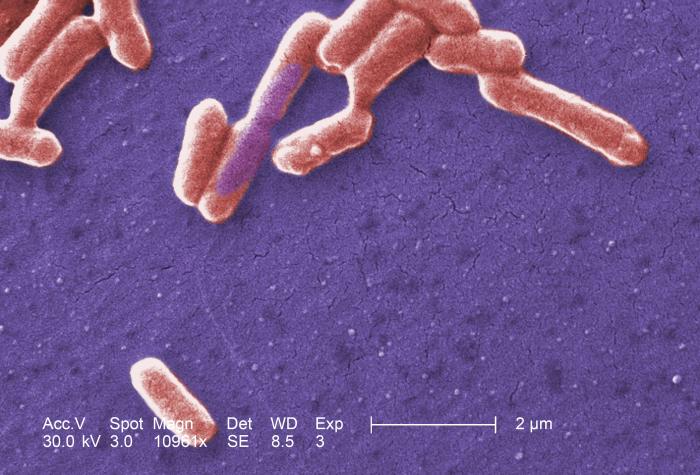
Colorized electron micrographs are useful illustrations, but the colors are often added for clarity and do not show a bacterium’s natural appearance. Magnification alone can also be deceptive if a scale bar is missing. Two photographs taken at different scales may make a narrow rod and a much larger cell look similar. Sample preparation can shrink or distort structures, and an image may show only one moment in a dynamic growth cycle. Readers should look for the organism’s name, imaging method, scale and preparation notes before treating a striking picture as evidence of how that organism behaves in its normal environment.
Put names beside real organisms
Escherichia coli is commonly rod-shaped, Staphylococcus aureus forms roughly spherical cells often seen in clusters, and Vibrio cholerae has a curved-rod form. These examples make the standard vocabulary useful, but even within a species cells can vary with growth conditions or stress. The organization of cells after division also matters: two spheres attached as a pair describe an arrangement, not a new basic cell shape. A microscope image without a scale bar can mislead because magnification alone does not tell viewers the true dimensions of the organisms. [1]
What cell-wall disruption can reveal
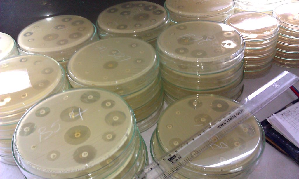
Bacterial shape is maintained through constant work, not by a wall that stays unchanged forever. As a rod-shaped bacterium grows, proteins direct where new wall material is inserted and where existing bonds are remodeled. Antibiotics that interfere with wall assembly can expose this dependence, producing elongation, distorted outlines or cell rupture in susceptible bacteria under particular conditions.
An experiment that changes cell-wall synthesis does not reveal one universal shape response. The outcome depends on the species, growth phase, osmotic environment and which molecular step is affected. Microscopy paired with viability measurements helps distinguish cells that have changed shape temporarily from cells that have lost the ability to grow. That is a more useful conclusion than assuming every elongated cell is dying.
References
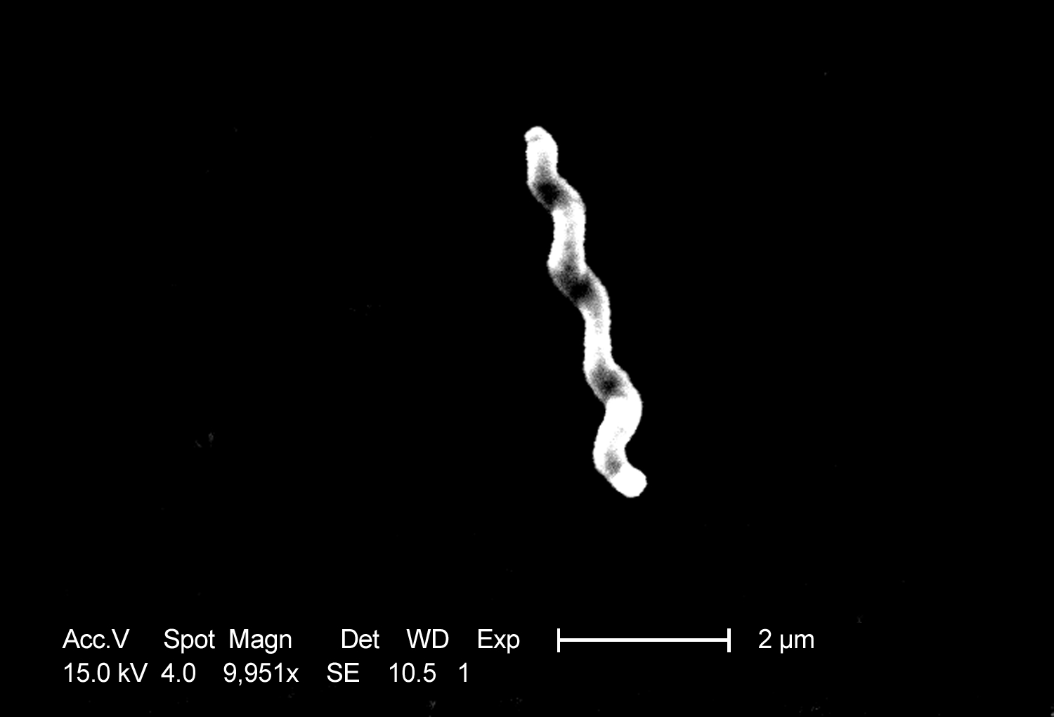
Featured image: Centers for Disease Control and Prevention, Public domain. Shows one curved bacterial morphology