A thin film on a tooth, a coating inside a pipe and microorganisms attached to a medical device can share one important feature: the cells live in a community fixed to a surface. These biofilms are not simply loose bacteria covered in glue. Their three-dimensional structure, physiology and local chemistry alter how cells obtain nutrients and respond to stress. Those details explain why removing them is often harder than treating free-floating cells.
A community attached to a surface
A biofilm begins when microorganisms attach to a surface and develop into a community, often held together by a self-produced extracellular matrix. The matrix can contain polysaccharides, proteins, lipids and extracellular DNA, with composition depending on the species and surroundings. Biofilms grow on rocks, teeth, pipes, medical devices and many other surfaces. They are not synonymous with disease. Environmental biofilms play important roles in nutrient cycling, and biofilm communities can be useful in engineered systems. Problems arise when their persistence is undesirable, especially in clinical or industrial settings. The key distinction is that cells living together on a surface encounter a different physical and chemical environment from freely suspended cells studied in a flask.
Why treatments behave differently
Antimicrobial susceptibility tests commonly use microorganisms growing in relatively standardized laboratory conditions. A mature biofilm presents additional obstacles. The extracellular matrix can slow the movement or local effectiveness of some compounds, while cells deeper in the community may grow slowly or enter physiological states less susceptible to certain drugs. Chemical gradients create pockets with different oxygen and nutrient availability. These effects are not all interchangeable with genetic resistance. Resistance refers to heritable mechanisms that allow organisms to grow despite a drug, while tolerance and persistence describe other forms of survival under exposure. An effective response must consider all of these mechanisms rather than assume that a stronger dose always solves the problem.
Microbes influence one another
Biofilms can contain one species or many. Neighboring organisms may compete for nutrients, produce inhibitory compounds or exchange metabolic products. Some species build matrix that benefits others, while another species may consume oxygen and create a niche suited to low-oxygen organisms. Communication signals sometimes influence collective behaviors, but quorum sensing is not required for every biofilm. Studies differ by organism and environment, so there is no universal bacterial script for forming a community. In oral plaque, for example, changing food availability and local acidity can shift which organisms prosper. The important unit of study is often the community and its environment, not a single isolated microbe.
Removal depends on the setting
Cleaning a hard industrial surface, managing dental plaque and treating an infected medical implant are not the same problem. Mechanical disruption can remove material that a chemical alone struggles to reach, but the appropriate approach depends on the surface and risk. In clinical care, management may involve targeted antimicrobials, drainage, device assessment or other interventions determined by professionals. Readers should not improvise treatment based on an article about general biofilm biology. In industrial systems, designers can reduce problematic attachment by controlling flow, maintenance and material selection. Even after a visible biofilm is removed, residual organisms or a newly exposed surface may allow recolonization. Prevention and sustained maintenance matter as much as a one-time cleaning step.
How researchers investigate biofilms
Scientists compare cells growing in suspension with cells attached to surfaces, using microscopy, flow cells, metabolic assays and measurements of viable organisms. Fluorescent labels can reveal spatial patterns, while sequencing helps describe which organisms are present. Yet a picture of thick material does not tell us whether every cell inside is alive, and a molecular survey does not automatically reveal which species are active. Studies using artificially smooth laboratory materials may not reproduce the texture and fluid movement of a real medical device or pipe. A careful investigation therefore reports the growth conditions, surfaces, treatment exposure and methods used to judge success. Biofilms are difficult partly because their properties emerge from place and history, not from species identity alone.
What a treatment study must distinguish
A surface may look clean after treatment while viable microorganisms remain, and a surface may remain stained even after a large reduction in living cells. Researchers therefore use several measurements: microscopy for spatial structure, culture-based tests for organisms that grow under the chosen conditions, and methods that estimate living cells or metabolic activity. Each tool has blind spots. A biofilm can also regrow from a small surviving population if the environment remains suitable. For clinical materials, the consequences of persistence differ sharply from those of harmless environmental films, and treatment decisions cannot be generalized across settings. A review of biofilm-associated resistance and tolerance explains why matrix effects, metabolic state and genetic mechanisms need to be considered separately. “Hard to remove” is not a single mechanism or one universal laboratory measurement.
How flowing water changes a biofilm
A biofilm forming inside a water line experiences forces that are absent from a static laboratory well. Flow brings nutrients and oxygen toward its surface, but it can also detach cells or fragments of the matrix. Faster flow is not simply better or worse: its effects depend on channel geometry, the strength of attachment and how mature the community has become.
Researchers use flow cells to examine these processes under microscopy while controlling the supply of medium. A biofilm may become patchy, form streamers or periodically release clusters of cells. That released material matters because it can travel downstream and attach elsewhere. Results from a static dish should not automatically be generalized to pipes, dental surfaces or medical devices without considering flow and surface chemistry.
Two surfaces, two very different settings
Dental plaque is a familiar biofilm: bacteria attach to tooth surfaces, consume nutrients and interact with other microorganisms within a growing community. A bacterial community on a catheter presents a different problem because organisms may remain attached to a medical device while interacting with tissue and immune defenses. Both are surface-associated, but the species, available nutrients, shear forces and control strategies differ. Mechanical disruption plays a central role in removing dental plaque; a device-associated infection may require medical decisions that cannot be inferred from a laboratory dish. [1]
Tolerance is not the same thing as resistance
Antibiotic resistance typically refers to inherited or acquired properties that let a bacterium grow at drug concentrations that would inhibit susceptible cells. Biofilm-associated tolerance can instead arise when cells enter slower-growing states or when environmental conditions within the community alter drug action. A biofilm may contain resistant cells, tolerant cells, or both. Researchers distinguish these possibilities by measuring survival and regrowth under controlled conditions, rather than assuming that any surviving attached bacteria carry a resistance mutation. This distinction shapes how experiments are reported and interpreted.
Some biofilm behaviors depend on chemical signals, but there is no single signaling system shared by every species. The distinction is developed in How Bacteria Communicate.
References
- Biofilm antibiotic resistance and tolerance review
- Ciofu et al.: Tolerance and resistance of microbial biofilms
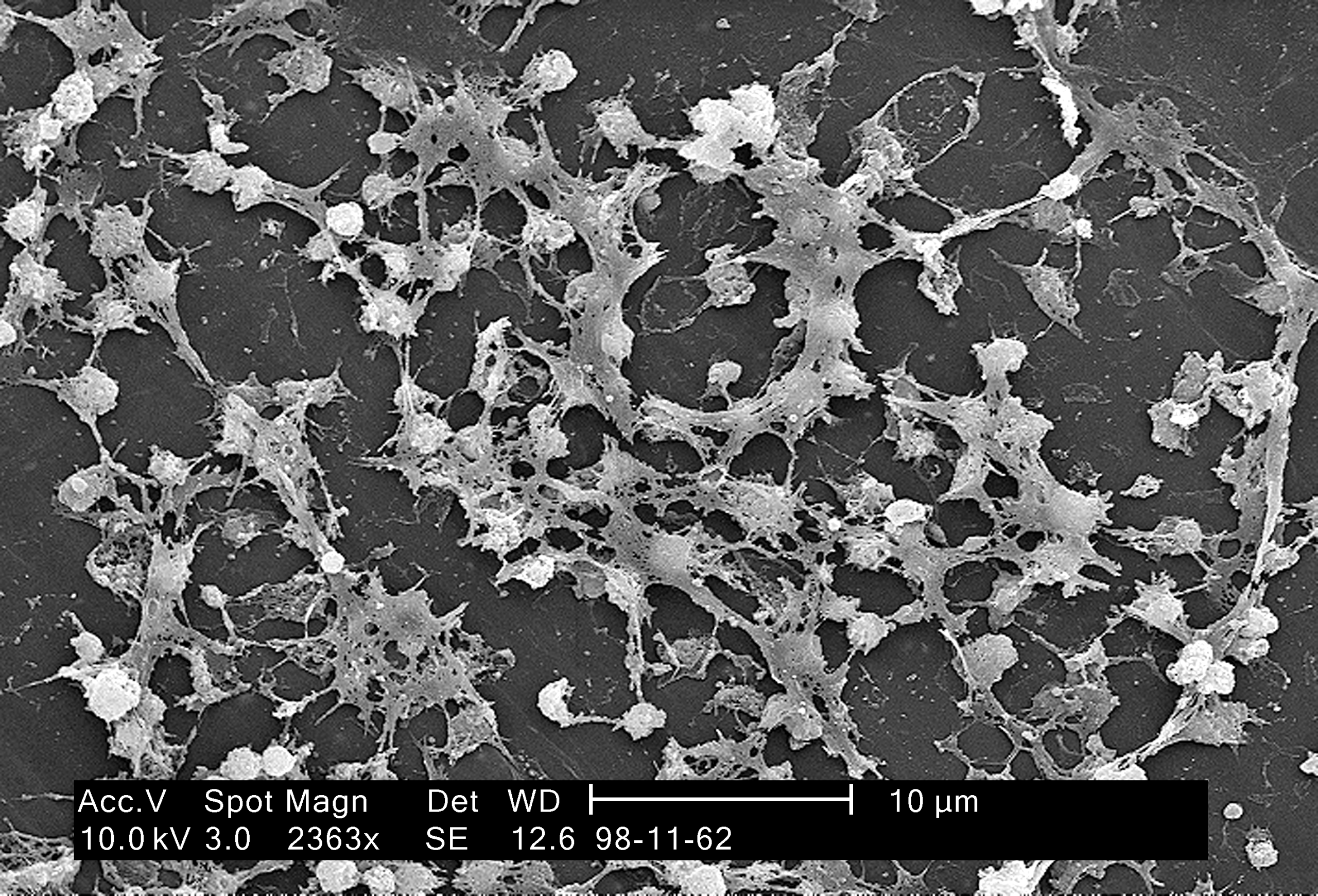
Featured image: CDC / Janice Carr, Public domain. Actual catheter-associated biofilm